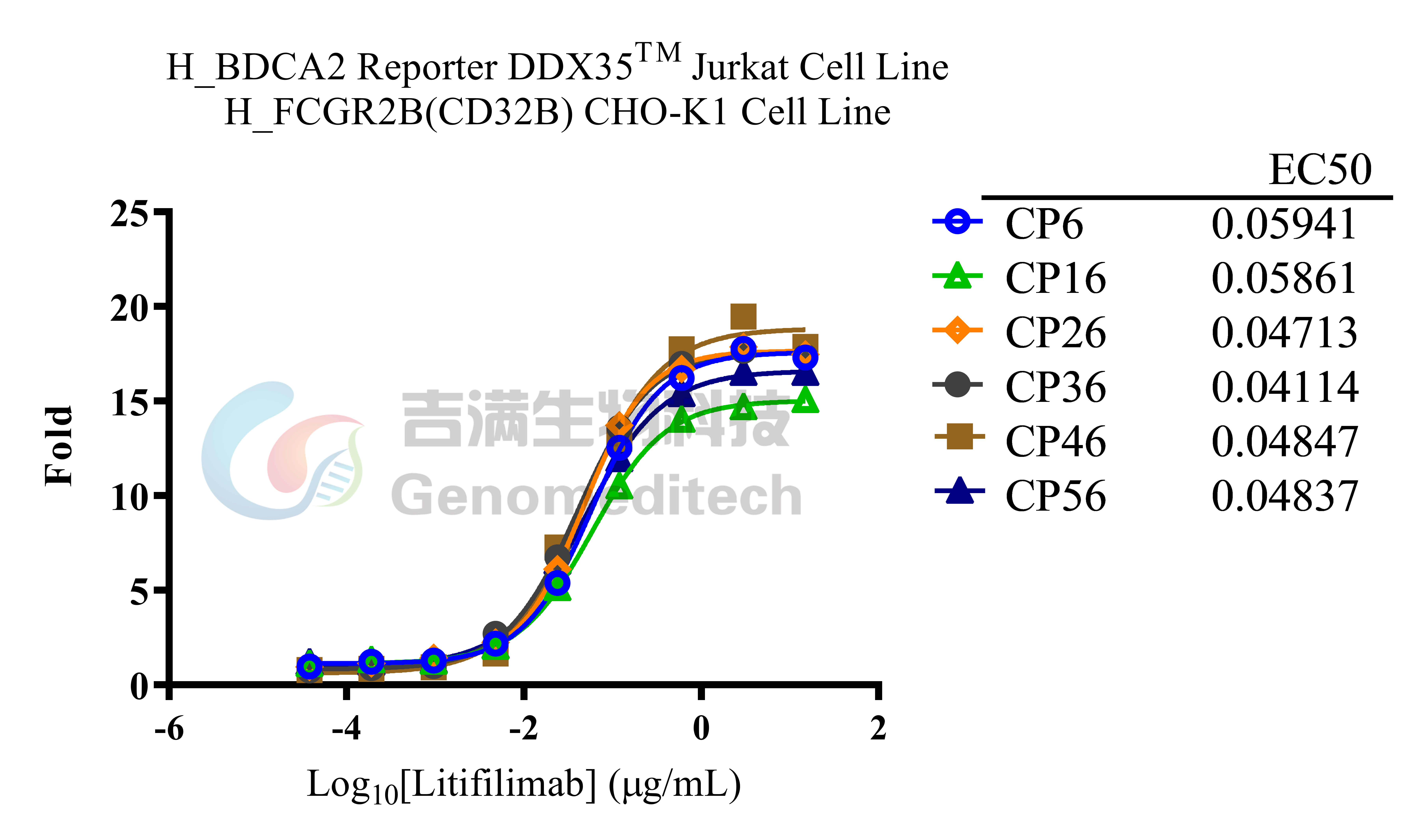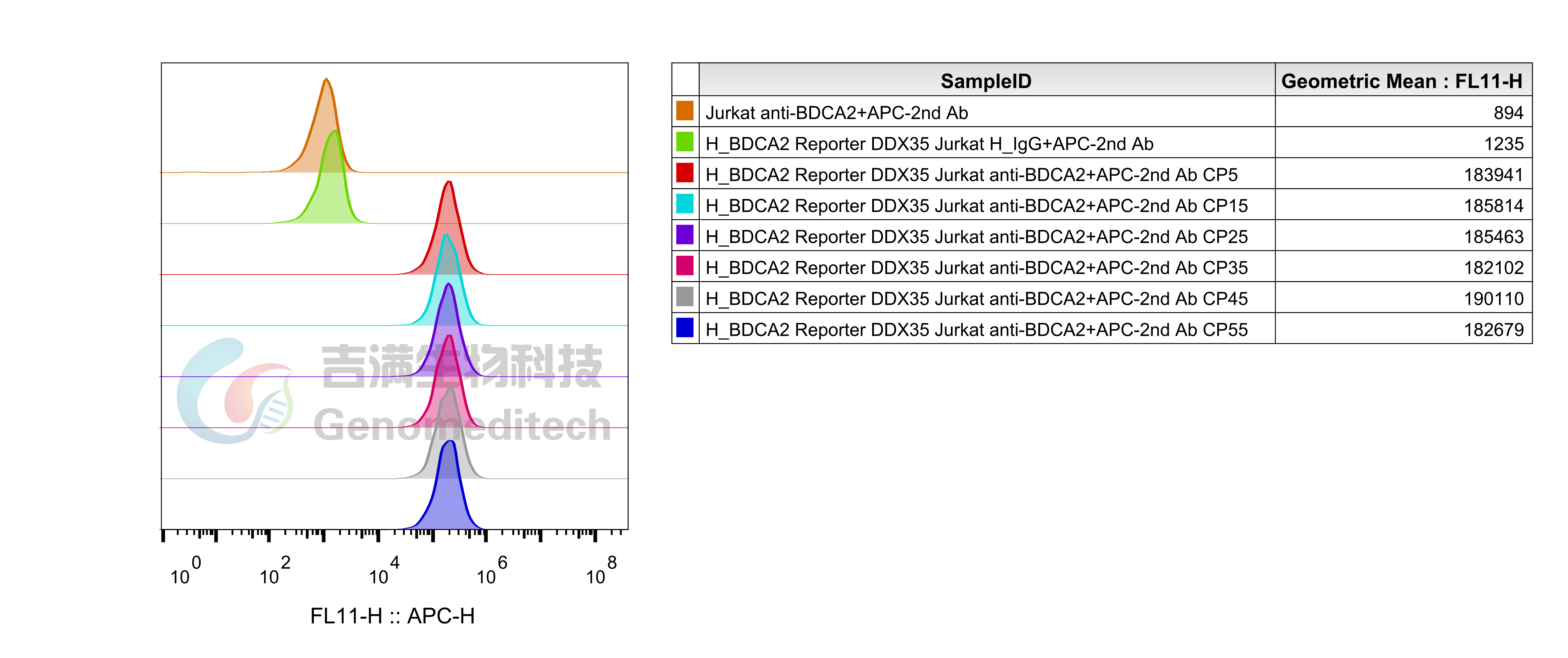


| Cat. No: GM-C39599 Product: H_BDCA2 Reporter DDX35TM Jurkat Cell Line Blood dendritic cell antigen 2 (BDCA2) is a C-type lectin expressed on plasmacytoid dendritic cells (pDCs) and is implicated in lupus pathogenesis. It consists of a single C-terminal extracellular carbohydrate recognition domain (CRD) of the class II C-type lectin family, a transmembrane region, and a short N-terminal cytoplasmic tail lacking a signaling motif. BDCA2 signals via the associated transmembrane adaptor FcεRIγ, triggering B cell receptor (BCR)-like signaling cascades. However, the ability of humanized anti-BDCA2 monoclonal antibodies to reduce disease activity in patients with cutaneous lupus remains poorly characterized. H_BDCA2 Reporter DDX35™ Jurkat Cell Line is a clonal stable Jurkat cell line constitutively expressing human BDCA2 and FcεRIγ gene, along with signal-dependent expression of a luciferase reporter gene. When drug stimulation is applied, it activates downstream signaling pathways, leading to the expression of luciferase. The measurement of luciferase activity indicates the activation level of the signaling pathway and can thus be used to evaluate the in vitro effects of an antibody targeting BDCA2. H_BDCA2 Reporter DDX35™ Jurkat Cell Line was obtained through extensive monoclonal screening and multiple rounds of monoclonal selection. It possesses high stability, high sensitivity, and high amplification properties, meeting the standards for customers' batch library construction and release experiments. | 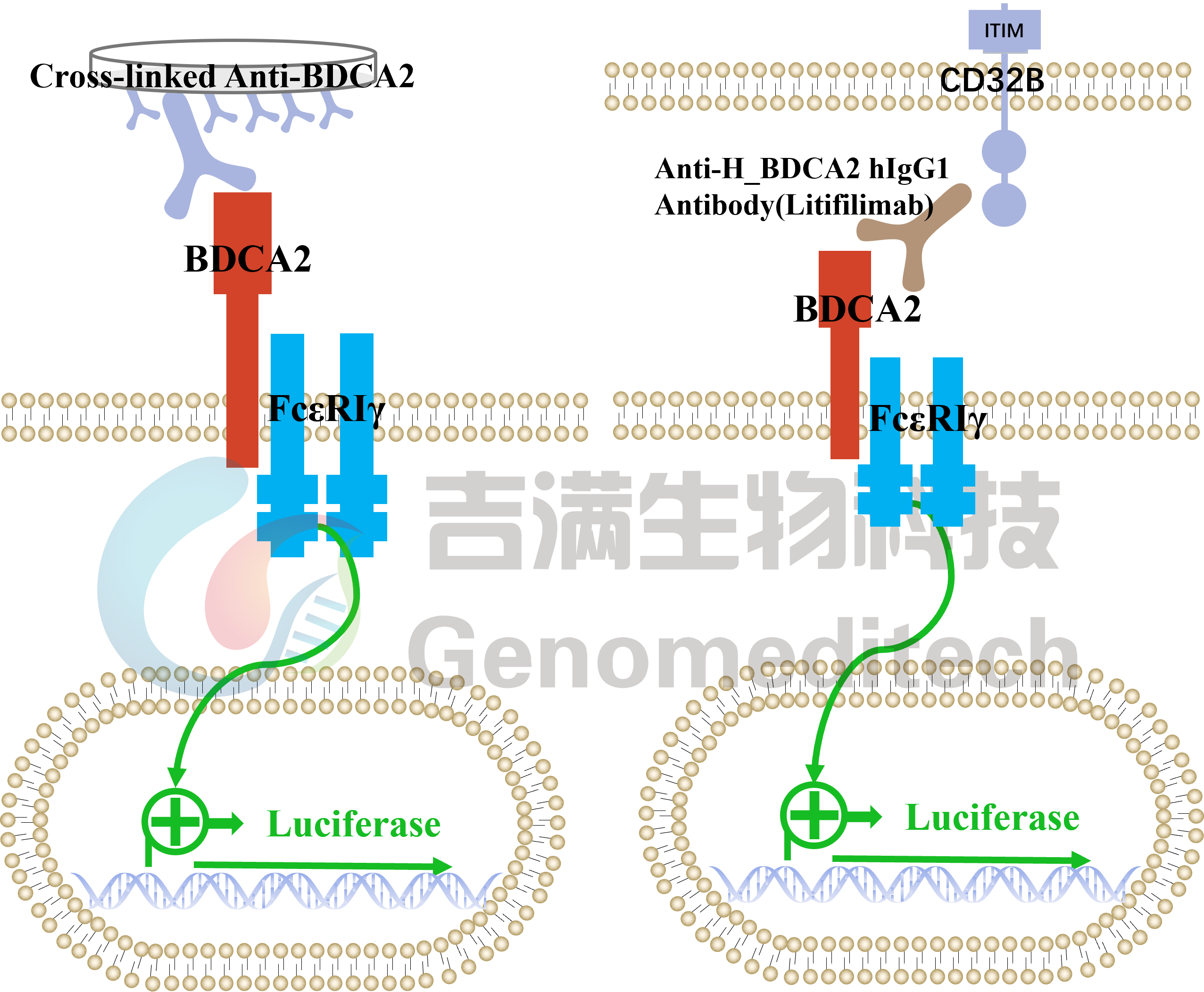 |