










PD-1 signaling represents a rare dual-directional target in biopharmaceutical drug development. On one side, PD-1/PD-L1 immune checkpoint inhibitors have achieved broad commercial success, with more than 20 related drugs approved worldwide, fundamentally reshaping the landscape of cancer immunotherapy. On the other, PD-1 agonists have emerged as a promising and increasingly active research area in autoimmune disease, aiming to reactivate PD-1-mediated inhibitory signaling and suppress hyperactivated autoreactive T cells, potentially addressing some of the safety limitations associated with conventional corticosteroids and JAK inhibitors.
However, whether developing a PD-1 inhibitor or agonist, biological activity assessment remains a critical checkpoint throughout the long journey from candidate molecule to marketed drug. It is not only a key parameter for CMC quality control, but also an essential component of the data package supporting IND/NDA submissions.
Therefore, a standardized in vitro model capable of evaluating both blocking activity and potential Fc-mediated agonistic effects has become increasingly important for accelerating PD-1 drug development.
New Product Launch: One Cell Line for PD-1 Blockade & Agonism
To further support drug development targeting the PD-1 pathway, Genomeditech H_PD-1 Reporter (IL-2 promoter) Jurkat Cell Line (GM-C43866) was developed.
Based on the human Jurkat T-cell line, this cell line was generated using lentiviral technology to establish a stable clone with constitutive PD-1 expression and an IL-2 promoter-driven luciferase reporter gene. Its key advantage is its dual functionality. By simply switching the co-culture partner, the same cell line can be used to establish functional evaluation models for both PD-1 blockade and PD-1 agonism:
PD-1 Blockade Mode
Co-culture with PD-L1-expressing target cells leads to PD-1/PD-L1 engagement and inhibition of TCR signaling, resulting in a decreased luciferase signal. Following the addition of a blocking antibody, this inhibitory effect is relieved and the signal is restored. A stronger signal indicates stronger blocking activity.
PD-1 Agonism Mode
Co-culture with FcγRIIb (CD32B)-expressing aAPC cells enables agonistic antibodies to promote PD-1 crosslinking through FcγRIIb. This enhances SHP2-mediated inhibitory signaling and further suppresses TCR activation, resulting in a decreased luciferase signal. A weaker signal indicates stronger agonistic activity.
This dual-use design eliminates the need to establish, maintain, and validate separate cell lines for PD-1 blockade and agonism, significantly reducing assay development costs and simplifying experimental system maintenance.
Why Choose the IL-2 Promoter?
Limitations of NFAT-Only Reporter Systems
Most commercially available PD-1 reporter cell lines use NFAT response elements to drive the reporter gene. Genomeditech takes a different approach by using the IL-2 promoter. This design is not simply an alternative choice, but is based on the underlying biology of PD-1 signaling.
NFAT is indeed a key transcription factor downstream of TCR signaling. However, PD-1 does not mediate its inhibitory effects through the NFAT pathway alone.
Following ligand engagement, PD-1 recruits the SHP2 phosphatase, which dephosphorylates multiple proximal signaling molecules, including CD28, PI3K, and ZAP70. This subsequently suppresses the PI3K/Akt/mTOR axis and ultimately reduces IL-2 gene transcription.
This means:
NFAT-only reporter gene systems capture only one branch of the inhibitory output downstream of PD-1. They can indicate that PD-1-mediated inhibition has occurred, but may not fully reflect the overall magnitude of PD-1-mediated suppression.
An IL-2 promoter-driven reporter, in contrast, measures the integrated output of the TCR-to-IL-2 signaling axis, incorporating signals mediated by NFAT, AP-1, NF-κB, and other transcriptional regulators. This provides a more comprehensive readout of the downstream functional consequences of PD-1 signaling.
Key Advantages of the IL-2 Promoter
The IL-2 gene promoter serves as a natural integration point for T-cell activation signals. It contains binding sites for NFAT, AP-1, NF-κB, and Oct-1, allowing it to respond to the integrated activity of multiple signaling pathways rather than a single transcription factor.
When PD-1 engages its ligand and recruits SHP2, the resulting inhibitory effects converge on IL-2 transcription. As IL-2 represents a key functional output of T-cell activation, an IL-2 promoter-driven reporter can provide an integrated readout of PD-1-mediated inhibition across multiple downstream signaling pathways.
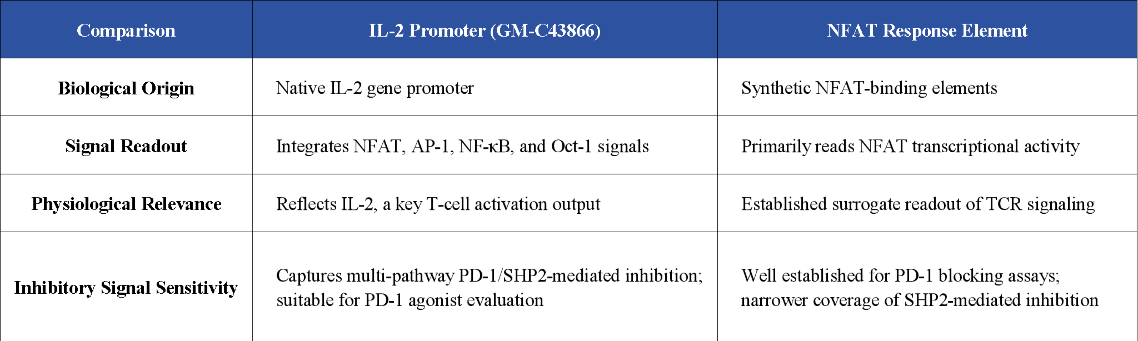
In short, the IL-2 promoter reporter system not only detects activation of PD-1 pathway signaling, but also quantitatively captures the extent of PD-1-mediated suppression of T-cell function, providing a physiologically relevant functional readout for potency assessment.
With this feature, the assay offers a broader dynamic response range and improved dose-response curve fitting, providing a reliable data foundation for EC50/IC50 determination, relative potency assessment, and drug comparability studies.
Three Key Application Scenarios and Experimental Data
This cell line can be paired with three complementary co-culture systems, covering the core functional evaluation needs in PD-1 drug development:
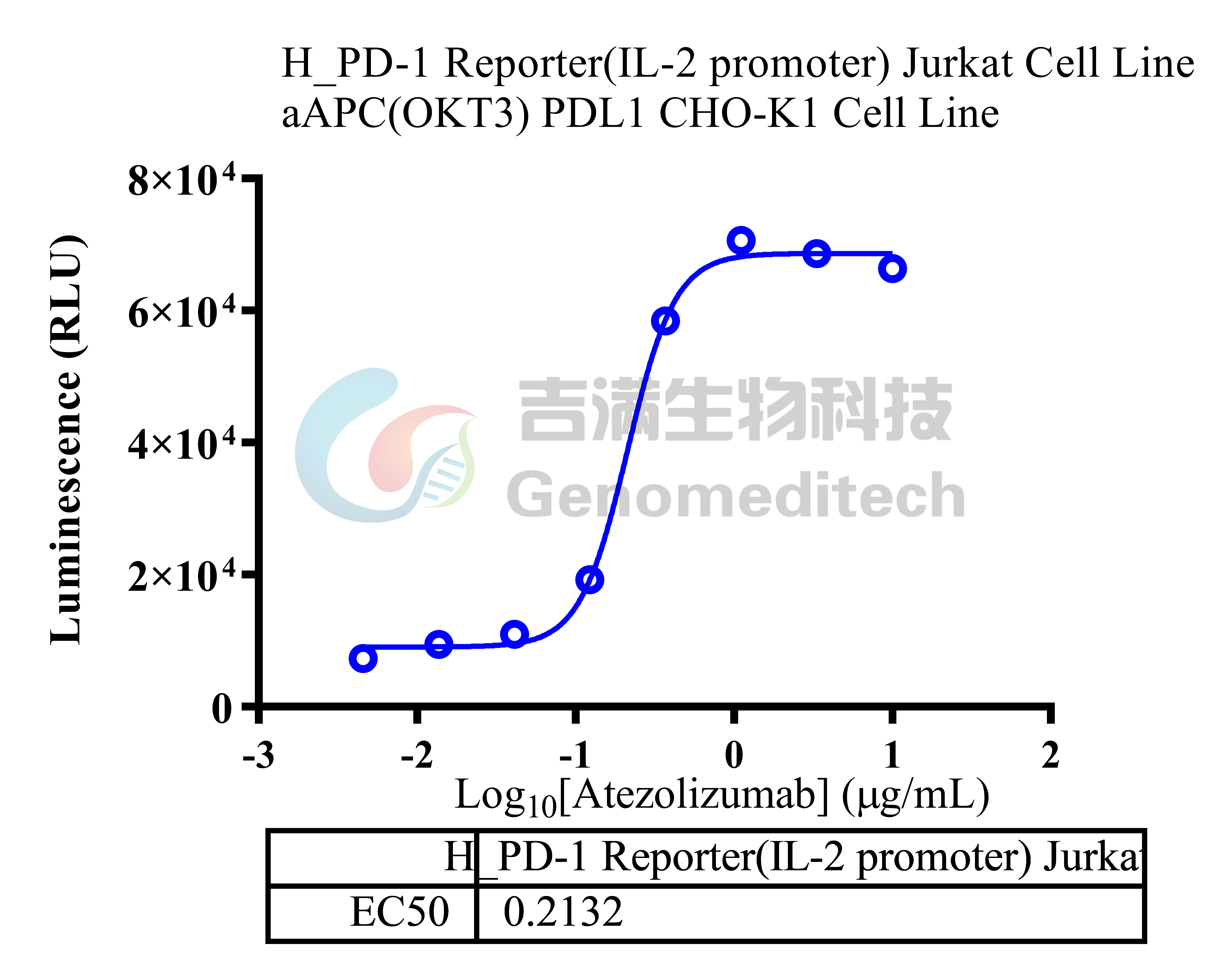
Scenario 1: Anti-PD-L1 Blocking Antibody Activity Evaluation
Co-culture Partner: aAPC (OKT3) PDL1 CHO-K1 Cell Line (GM-C05269)
Reference Antibody: Anti-H_PDL1 hIgG1 Reference Antibody (GM-86854MAB)
Experimental Procedure: aAPC cells were plated overnight. On the following day, serially diluted test antibodies were co-incubated with reporter cells for 1 h, followed by 16 h of co-culture in the assay plate. Luciferase activity was then measured.
Maximum response fold change ≈ 11.9-fold
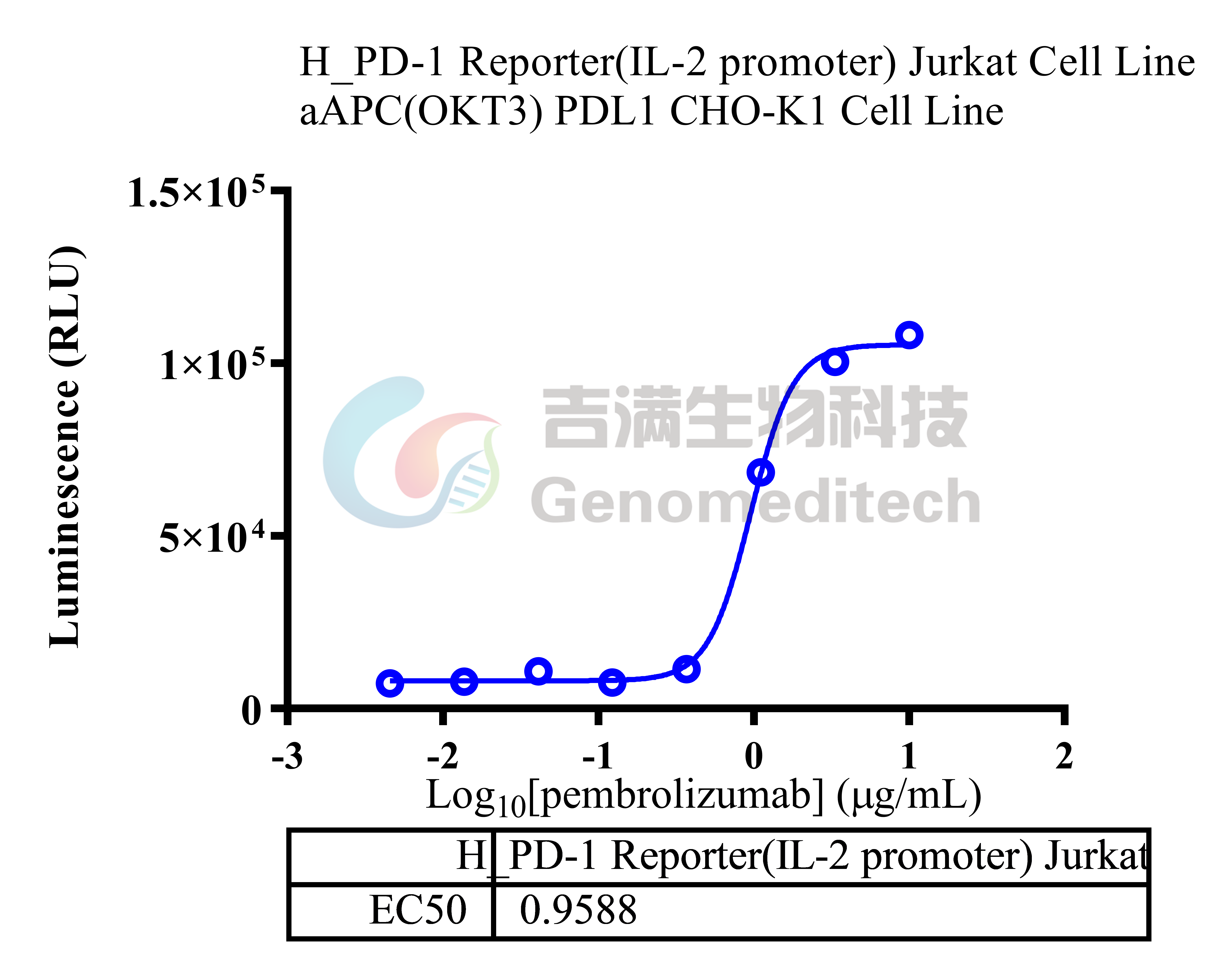
Scenario 2: Anti-PD-1 Blocking Antibody Activity Evaluation
Co-culture Partner: aAPC (OKT3) PDL1 CHO-K1 Cell Line (GM-C05269)
Reference Antibody: Anti-PD1 hIgG4 Reference Antibody (GM-87802MAB)
Experimental Procedure: Same as above, with an anti-PD-1 antibody used in place of the anti-PD-L1 antibody.
Maximum response fold change ≈ 13.3-fold
Scenario 3: PD-1 Agonist Antibody Potency Evaluation
Co-culture Partner: H_CD32B aAPC CHO-K1 Cell Line (GM-C25754)
Principle: PD-1 agonist antibodies promote crosslinking of PD-1 with the FcγRIIb receptor, activating SHP2-mediated inhibitory signaling downstream of PD-1 and suppressing TCR activation, resulting in a decreased luciferase signal.
Tested Antibodies: Anti-PDl hlgG1 Reference Antibody
Maximum response fold change ≈ 25.5-fold
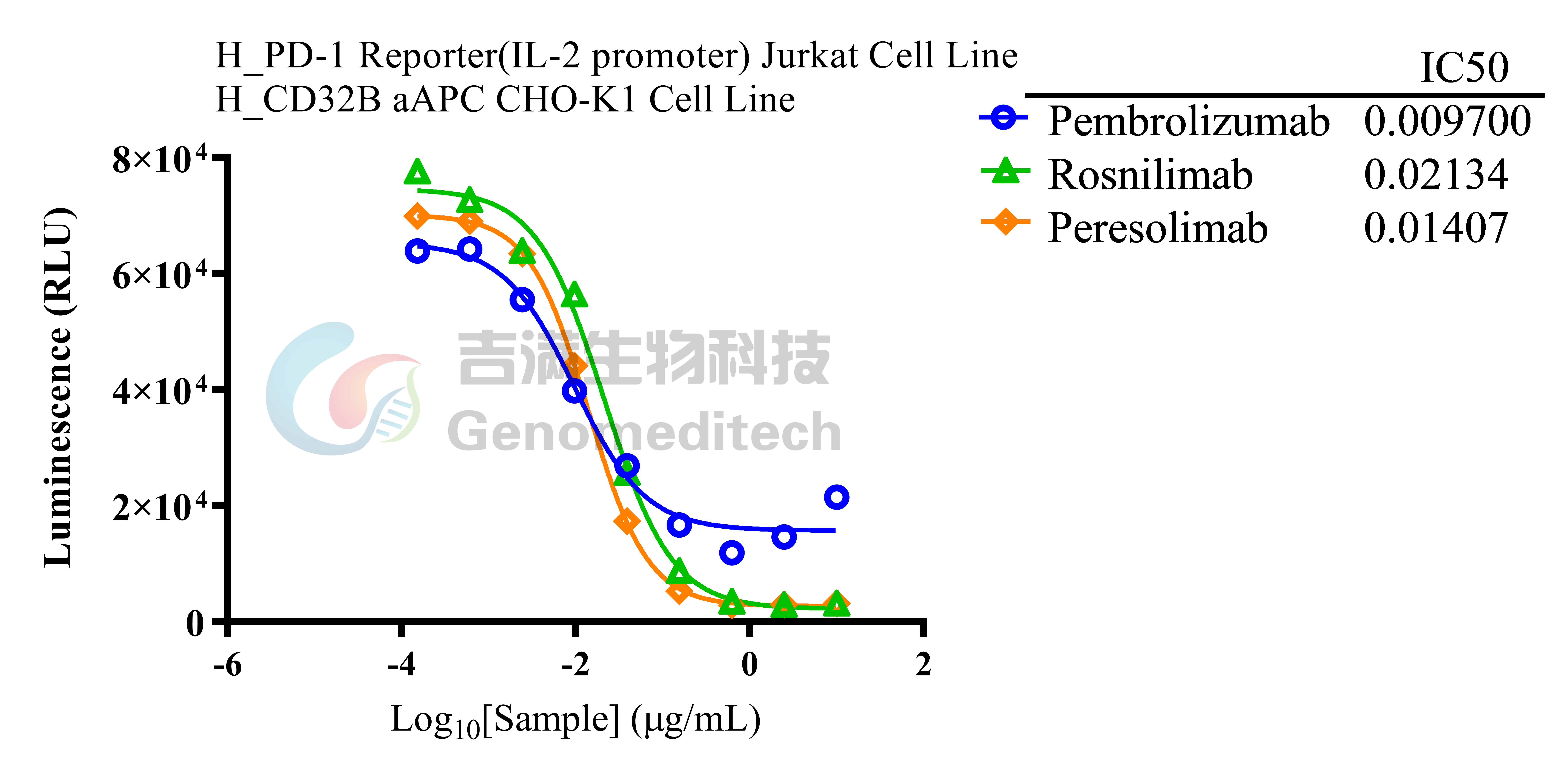
These data demonstrate that whether developing PD-1/PD-L1 blocking antibodies for oncology applications or PD-1 agonists for autoimmune disease, GM-C43866 provides a sufficiently broad response window and reliable dose-response curves. This makes it suitable for functional activity assessment throughout the drug development process, from early-stage screening to QC and lot release.
Industrial-Grade Quality: Built for Compliance
For a cell line to support IND/NDA submissions, meeting laboratory-use requirements is only the starting point. Genomeditech has established a comprehensive quality and compliance framework:
Global Commercial Use Authorization
Genomeditech holds global commercial use authorization for HEK293, Jurkat E6.1, and CHO-K1 cell lines, providing a clear IP and compliance foundation for drug development and regulatory submissions across major markets.
ISO 9001 Quality System
Standardized quality control is implemented throughout cell line development, expansion, and release testing, including functional validation and mycoplasma testing.
Passage Stability Validation
Consistent assay performance has been validated across P4, P14, and P24 passages, supporting long-term QC applications and cross-site method transfer.
Complete Product Ecosystem
Complementary co-culture cell lines (GM-C05269, GM-C03541, and GM-C25754), reference antibodies, and the GMOne-Step 2.0 Luciferase Reporter Gene Assay Kit provide an integrated solution for PD-1 functional evaluation.
Scale and Coverage
Nearly 400 targets and more than 1,500 off-the-shelf monoclonal cell lines support consistent supply and rapid delivery for global drug development programs.
Product Overview
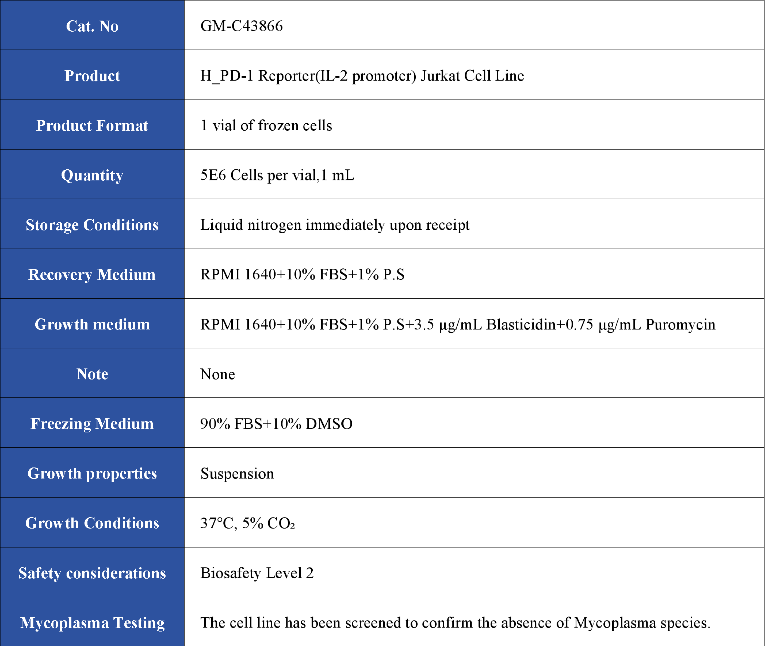
Learn More: https://en.genomeditech.com/cell-line/H_PD-1-Reporter%28IL-2-promoter%29-Jurkat-Cell-Line